The art-labeling activity: structure and bands of the sarcomere embarks on an illuminating journey into the intricate realm of muscle biology. This comprehensive guide delves into the fundamental organization of the sarcomere, the basic unit of muscle contraction, unraveling its structural components and their functional significance with unparalleled clarity and precision.
Through detailed diagrams, informative tables, and engaging visuals, this activity provides a thorough understanding of the sarcomere’s architecture, including the arrangement of myofilaments and Z-lines. It meticulously identifies the distinct bands visible under a microscope, exploring their composition and function, such as the A-band, I-band, H-zone, and M-line.
Structure of the Sarcomere
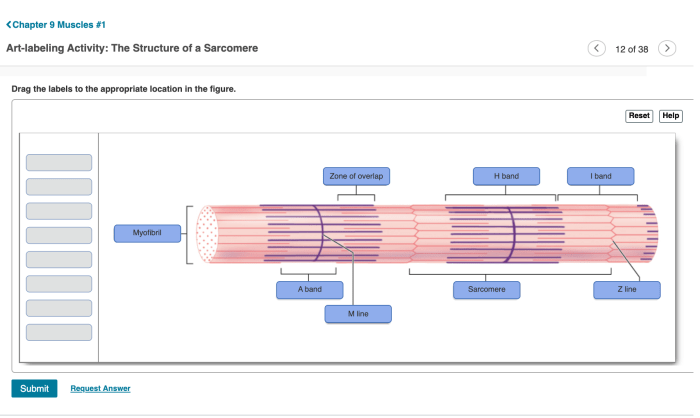
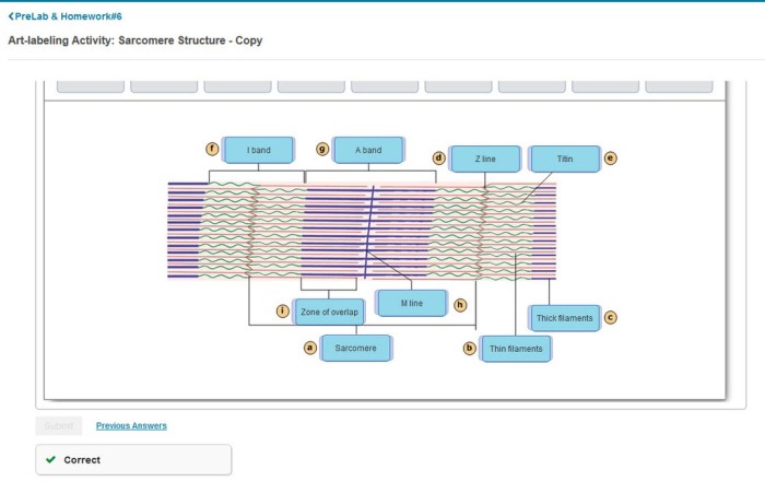
The sarcomere is the basic unit of muscle contraction. It is composed of two types of myofilaments: thick myosin filaments and thin actin filaments. The myofilaments are arranged in a repeating pattern within the sarcomere, creating the characteristic banding pattern visible under a microscope.
Arrangement of Myofilaments
Myosin filaments are located in the center of the sarcomere, while actin filaments are located on either side of the myosin filaments. The actin filaments are attached to the Z-lines, which are dense structures that run across the sarcomere. The myosin filaments are not attached to the Z-lines, but instead project into the space between the actin filaments.
Regions of the Sarcomere, Art-labeling activity: structure and bands of the sarcomere
The sarcomere is divided into several distinct regions based on the arrangement of the myofilaments:
- A-band: The A-band is the central region of the sarcomere and contains both myosin and actin filaments.
- I-band: The I-band is the region on either side of the A-band and contains only actin filaments.
- H-zone: The H-zone is the central region of the A-band and contains only myosin filaments.
- M-line: The M-line is a thin line in the center of the H-zone and is thought to help stabilize the myosin filaments.
Bands of the Sarcomere

The different bands visible in a sarcomere under a microscope are:
A-band
The A-band is the central region of the sarcomere and contains both myosin and actin filaments. It is the thickest band in the sarcomere and is responsible for the muscle’s ability to contract.
I-band
The I-band is the region on either side of the A-band and contains only actin filaments. It is the thinnest band in the sarcomere and is responsible for the muscle’s ability to relax.
H-zone
The H-zone is the central region of the A-band and contains only myosin filaments. It is the region where the myosin heads interact with the actin filaments to cause muscle contraction.
M-line
The M-line is a thin line in the center of the H-zone and is thought to help stabilize the myosin filaments.
Organization of Myofilaments: Art-labeling Activity: Structure And Bands Of The Sarcomere

Myosin and actin filaments are arranged in a specific way within the sarcomere. Myosin filaments are located in the center of the sarcomere, while actin filaments are located on either side of the myosin filaments. The actin filaments are attached to the Z-lines, which are dense structures that run across the sarcomere.
The myosin filaments are not attached to the Z-lines, but instead project into the space between the actin filaments.The myosin and actin filaments are arranged in a repeating pattern, creating the characteristic banding pattern visible under a microscope. The A-band is the central region of the sarcomere and contains both myosin and actin filaments.
The I-band is the region on either side of the A-band and contains only actin filaments. The H-zone is the central region of the A-band and contains only myosin filaments. The M-line is a thin line in the center of the H-zone and is thought to help stabilize the myosin filaments.
Sliding Filament Model of Muscle Contraction

The sliding filament model of muscle contraction explains how the myofilaments interact to cause muscle contraction. According to this model, the myosin filaments slide past the actin filaments, causing the sarcomere to shorten.The sliding filament model is initiated by the release of calcium ions from the sarcoplasmic reticulum.
The calcium ions bind to receptors on the troponin complex, which is located on the actin filaments. This causes the troponin complex to change shape, exposing the myosin-binding sites on the actin filaments.The myosin heads then bind to the myosin-binding sites on the actin filaments, forming cross-bridges.
The myosin heads then pivot, pulling the actin filaments towards the center of the sarcomere. This causes the sarcomere to shorten and the muscle to contract.The sliding filament model is a repeating process that continues until the calcium ions are removed from the sarcoplasmic reticulum.
When the calcium ions are removed, the troponin complex changes shape again, blocking the myosin-binding sites on the actin filaments. This causes the myosin heads to detach from the actin filaments and the muscle to relax.
Functional Significance of Sarcomere Structure
The structure of the sarcomere is directly related to its function. The arrangement of the myofilaments and the different bands within the sarcomere allow for efficient muscle contraction.The thick myosin filaments in the center of the sarcomere provide the power for muscle contraction.
The thin actin filaments on either side of the myosin filaments provide the surface for the myosin heads to bind to. The Z-lines anchor the actin filaments in place and help to maintain the proper alignment of the myofilaments.The different bands within the sarcomere also play important roles in muscle contraction.
The A-band is the region where the myosin heads interact with the actin filaments to cause muscle contraction. The I-band is the region where the actin filaments are not overlapped by myosin filaments, allowing for muscle relaxation. The H-zone is the central region of the A-band where the myosin heads are not attached to the actin filaments.
This region allows for the myosin heads to pivot and pull the actin filaments towards the center of the sarcomere.The structure of the sarcomere is essential for efficient muscle contraction. The arrangement of the myofilaments and the different bands within the sarcomere allow for the generation of force and the control of muscle contraction.
FAQ Resource
What is the significance of the Z-line in sarcomere structure?
The Z-line serves as the anchor point for actin filaments, providing structural stability to the sarcomere and facilitating the transmission of force during muscle contraction.
How does the sliding filament model explain muscle contraction?
The sliding filament model proposes that muscle contraction occurs when myosin filaments slide past actin filaments, shortening the sarcomere and generating force.
What factors influence the force generated by a muscle?
The force generated by a muscle is influenced by factors such as the number of sarcomeres in parallel, the length of the sarcomeres, and the availability of calcium ions.