Laryngoscope size for 32 weeks is a crucial aspect of fetal intubation, influencing the success and safety of the procedure. Understanding the anatomical considerations, laryngoscope blade selection, and proper insertion technique is paramount. This guide will delve into these key elements, providing a comprehensive overview for healthcare professionals.
As fetal development progresses, the anatomy of the airway undergoes significant changes. At 32 weeks, the larynx is located higher in the neck, and the epiglottis is more pliable, necessitating specific laryngoscope blade sizes and shapes to ensure optimal visualization and minimize trauma.
Anatomical Considerations: Laryngoscope Size For 32 Weeks
Selecting the appropriate laryngoscope size for a 32-week fetus requires an understanding of the relevant anatomical structures and their developmental changes during pregnancy.
The laryngoscope is inserted into the oropharynx and advanced along the posterior pharyngeal wall until the epiglottis is visualized. The epiglottis is a leaf-shaped cartilage that covers the entrance to the larynx during swallowing.
Developmental Changes
As the fetus develops, the epiglottis and other laryngeal structures increase in size and change in shape. At 32 weeks, the epiglottis is typically approximately 10-12 mm in length and is more horizontal than in adults.
Laryngoscope Blade Selection

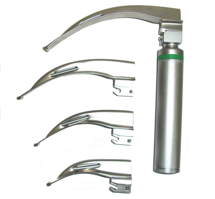
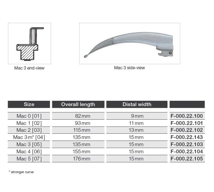
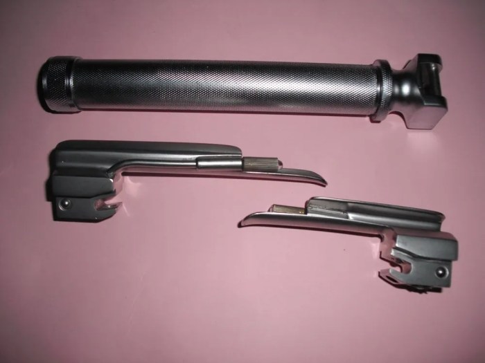
Laryngoscope Blade Sizes and Shapes for 32-Week Fetuses
The selection of the appropriate laryngoscope blade size and shape is crucial for successful intubation of 32-week fetuses. The size and shape of the blade must be carefully matched to the fetal anatomy to minimize trauma and ensure effective visualization of the glottis.
Blade Selection Rationale
The choice of laryngoscope blade is based on several factors, including the fetal head circumference, gestational age, and anticipated difficulty of intubation. For 32-week fetuses, the following guidelines apply:
- Head circumference:The head circumference is a good indicator of the overall size of the fetus. A larger head circumference may require a larger blade size.
- Gestational age:As the fetus grows, the size of the airway increases. Therefore, older fetuses may require larger blade sizes.
- Anticipated difficulty of intubation:If the intubation is expected to be difficult, a smaller blade size may be preferred to avoid excessive force and trauma.
Blade Size and Shape Comparison
The following table compares different laryngoscope blade sizes and shapes for 32-week fetuses:
| Blade Size | Blade Shape | Suitability for 32-Week Fetuses |
|---|---|---|
| Miller 0 | Straight | Suitable for most 32-week fetuses |
| Miller 1 | Straight | May be required for larger 32-week fetuses |
| Macintosh 1 | Curved | Suitable for smaller 32-week fetuses |
| Macintosh 2 | Curved | May be required for larger 32-week fetuses |
Insertion Technique

Inserting a laryngoscope in 32-week fetuses requires meticulous technique to optimize visualization and minimize trauma. Here’s a detailed guide to the proper insertion procedure.
Blade Placement:
- Gently insert the laryngoscope blade along the fetal mouth’s midline, avoiding the tongue and epiglottis.
- Advance the blade until the tip rests just below the epiglottis, forming a slight angle with the fetal spine.
Vocal Cord Visualization:
- Lift the blade gently upwards and anteriorly, creating a clear view of the vocal cords.
- Maintain gentle pressure to stabilize the blade while visualizing the vocal cords and surrounding structures.
Tips for Optimization and Minimization of Trauma:
- Use the smallest blade size possible to minimize tissue damage.
- Insert the blade slowly and cautiously, avoiding any sudden movements.
- Apply minimal pressure to the blade to prevent excessive force on delicate tissues.
- Maintain a clear view of the vocal cords to avoid inadvertent contact with other structures.
Alternative Laryngoscopy Methods

Alternative laryngoscopy methods offer additional options for visualizing the laryngeal structures of 32-week fetuses. These techniques provide distinct advantages and disadvantages, expanding the range of available approaches.
Videolaryngoscopy
Videolaryngoscopy employs a small camera mounted on the laryngoscope blade, providing a real-time video feed of the larynx on a monitor. Advantages:
- Enhanced visualization and magnification
- Improved ergonomics and reduced strain on the operator
- Ability to record and review procedures
Disadvantages:
- Requires specialized equipment and training
- May be more expensive than traditional laryngoscopy
Fiberoptic Laryngoscopy
Fiberoptic laryngoscopy utilizes a flexible fiberoptic cable with a camera at its tip, allowing for indirect visualization of the larynx. Advantages:
- Less invasive and traumatic
- Can navigate challenging airways
- Allows for prolonged visualization
Disadvantages:
For 32 weeks, the laryngoscope size is typically 3-4 mm. This size is appropriate for infants of this gestational age. However, for more information on biomolecules, you can check out hhmi biomolecules on the menu . Returning to our topic, it’s important to use the correct laryngoscope size for infants of different gestational ages to ensure proper visualization of the larynx.
- Requires specialized training and skill
- Can be time-consuming
- May provide a narrower field of view
Direct Laryngoscopy with Flexible Endoscope, Laryngoscope size for 32 weeks
This technique combines direct laryngoscopy with a flexible endoscope, inserted through the laryngoscope blade. Advantages:
- Combines the benefits of both methods
- Allows for direct visualization and manipulation
- Reduces trauma compared to rigid endoscopes
Disadvantages:
- Requires additional equipment and training
- Can be more challenging to use than videolaryngoscopy
Comparison of Alternative Laryngoscopy Methods
| Method | Indications | Contraindications ||—|—|—|| Videolaryngoscopy | Difficult airway, limited neck mobility, obese patients | Equipment failure, operator inexperience || Fiberoptic Laryngoscopy | Preterm infants, difficult airways, tracheal stenosis | Bleeding disorders, esophageal perforation || Direct Laryngoscopy with Flexible Endoscope | Failed videolaryngoscopy, challenging airways, need for airway manipulation | Significant airway trauma, esophageal perforation |
Safety Considerations

Ensuring patient safety is paramount during laryngoscopy in 32-week fetuses, given their delicate anatomy and vulnerability. Understanding the potential risks and complications associated with the procedure is crucial for minimizing harm and ensuring a successful outcome.
Potential Risks and Complications
- Premature rupture of membranes (PROM): The insertion of the laryngoscope can inadvertently puncture the amniotic sac, leading to PROM.
- Fetal bradycardia: The pressure applied during laryngoscopy can stimulate the vagus nerve, causing a decrease in fetal heart rate.
- Fetal injury: The laryngoscope blade can potentially cause trauma to the fetal face, airway, or vocal cords if not handled with utmost care.
- Infection: The introduction of instruments into the fetal airway carries the risk of infection, although this is rare.
Guidelines for Minimizing Risks
- Gentle insertion: Use the laryngoscope blade gently and avoid excessive force when inserting it into the fetal mouth.
- Proper positioning: Ensure the fetus is in an optimal position for laryngoscopy, with the head extended and the neck slightly flexed.
- Experienced operator: The procedure should be performed by an experienced and skilled practitioner who is familiar with fetal anatomy and laryngoscopy techniques.
- Adequate monitoring: Closely monitor the fetus during and after the procedure for any signs of distress or complications.
- Emergency preparedness: Be prepared to manage any potential complications that may arise, such as PROM or fetal bradycardia.
Top FAQs
What is the optimal laryngoscope blade size for 32-week fetuses?
The recommended blade size for 32-week fetuses is typically a Miller 0 or 1 blade.
How should the laryngoscope blade be inserted for 32-week fetuses?
The blade should be inserted gently into the vallecula, avoiding excessive pressure on the epiglottis or arytenoids.
What are the potential complications associated with laryngoscopy in 32-week fetuses?
Potential complications include airway trauma, laryngeal edema, and vocal cord damage. Careful technique and appropriate blade selection can minimize these risks.